


Pigmented Villonodular Synovitis (PVNS): Symptoms, Causes & Treatment
Abstract
Pigmented Villonodular Synovitis (PVNS), now commonly classified within the spectrum of tenosynovial giant cell tumors, is an uncommon proliferative disorder affecting the synovial lining of joints, bursae, and tendon sheaths. It is characterized by excessive synovial proliferation and hemosiderin accumulation, leading to progressive joint swelling, pain, stiffness, and restricted movement. The knee and hip are frequently involved, although other joints may also be affected. Magnetic resonance imaging is particularly valuable for identifying characteristic synovial changes and hemosiderin deposition, while histopathological examination confirms the diagnosis. Management depends on disease extent and may include arthroscopic or open synovectomy, with systemic therapies considered in selected diffuse cases. From an Ayurvedic perspective, its clinical manifestations may be considered in relation to disorders involving Vata-Kapha imbalance, Srotorodha (channel obstruction), and localized Dhatu involvement. An individualized approach emphasizing Dosha balance, Agni support, and appropriate external and internal therapies may provide supportive care alongside conventional management.
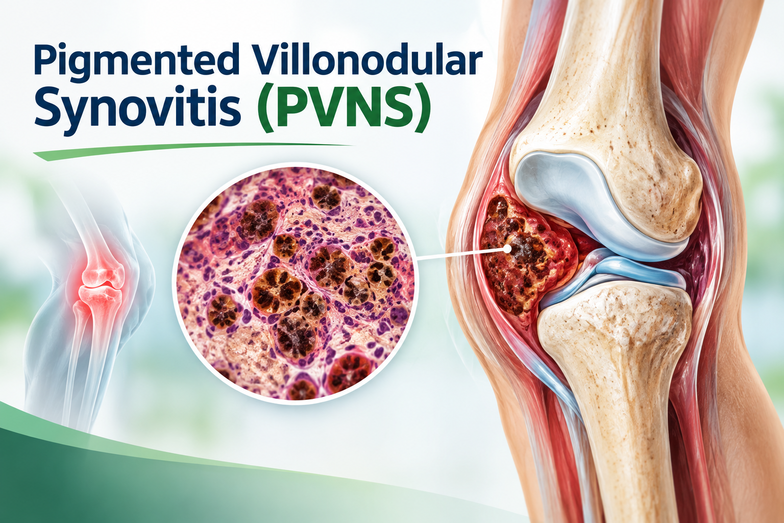
Statistics for Pigmented Villonodular Synovitis
- Pigmented villonodular synovitis (PVNS), currently included under the spectrum of tenosynovial giant cell tumors (TGCTs), is an uncommon benign synovial proliferative condition, with an estimated annual incidence of approximately 2–9.2 cases per million people.
- The disorder predominantly affects young and middle-aged adults, with most cases occurring between 20 and 50 years of age.
- A slight female predominance has been reported, with female-to-male ratios ranging from approximately 1.35:1 to 2:1 across different studies and disease subtypes.
- Studies have reported a median diagnostic interval of around 18 months following the onset of symptoms.
- Localized PVNS generally has a more favorable clinical course than the diffuse form. Following complete surgical excision, the reported recurrence rate is approximately 8.6–9%.
Interesting Facts about Pigmented Villonodular Synovitis
- It can “bleed” without an injury: PVNS often contains extensive hemosiderin, an iron-storage pigment produced from repeated microscopic bleeding within the affected synovium. This gives the lesion its characteristic brownish appearance.
- MRI can reveal its hidden identity: The hemosiderin deposits create a distinctive low-signal appearance on MRI, particularly on gradient-echo or susceptibility-sensitive sequences.
- It is not actually a tumor of the bone: Despite sometimes producing pressure erosions of adjacent bone, the primary abnormality originates from the synovial tissue, tendon sheath, or bursa.
- One disease, two very different behaviors: PVNS may appear as a localized nodular lesion or as a diffuse process involving extensive synovium. The diffuse form is substantially more difficult to control and has a greater tendency to recur.
- The knee is not its only home: Although the knee and hip are frequent sites, PVNS can occur in unusual locations, including the ankle, shoulder, wrist, elbow, and temporomandibular joint.
- Its name describes what the surgeon may see: “Pigmented” refers to hemosiderin deposition, “villonodular” describes the finger-like villous and nodular growth, and “synovitis” reflects involvement of the synovial lining.
- It can hide behind an ordinary joint complaint: A patient may initially have only intermittent swelling, discomfort, or restricted movement, allowing PVNS to remain undiagnosed for months or even years.
Introduction
Pigmented Villonodular Synovitis (PVNS) is an uncommon disorder of synovial tissue that presents an important diagnostic challenge in musculoskeletal medicine. The condition is characterized by abnormal expansion of synovial tissue within a joint or, less frequently, around tendon sheaths and bursae. Although its clinical behavior is generally benign, the progressive nature of the lesion can interfere substantially with joint function and, in longstanding cases, contribute to cartilage and subchondral bone damage. The disease is now understood within the broader classification of tenosynovial giant cell tumors (TGCTs), which encompasses localized and diffuse patterns of synovial involvement. Its underlying biology is linked to dysregulated colony-stimulating factor 1 (CSF1) signaling, which promotes recruitment and accumulation of macrophages and other inflammatory cells within the lesion. This cellular environment accounts for several of its characteristic pathological features, including multinucleated giant cells, lipid-laden macrophages, and hemosiderin deposition. Clinically, PVNS may present with persistent or recurrent joint swelling, pain, stiffness, reduced range of motion, or episodes resembling spontaneous hemarthrosis.
Causes of Pigmented Villonodular Synovitis
The exact cause of Pigmented Villonodular Synovitis (PVNS) is not completely established. Current evidence suggests that it develops primarily through abnormal genetic signaling within synovial tissue, rather than from a single external trigger.
- CSF1 Gene Abnormality – A major molecular feature in many cases is an abnormality involving the colony-stimulating factor 1 (CSF1) gene. Increased CSF1 production promotes the recruitment of numerous macrophages into the affected synovial tissue.
- CSF1–CSF1R Signaling Abnormality – CSF1 binds to its receptor, CSF1R, on macrophages and related cells. Excessive activation of this signaling pathway results in macrophage proliferation and accumulation.
- Chromosomal Rearrangements – Structural changes involving chromosome 1p13, where the CSF1 gene is located, have been identified in a proportion of PVNS/TGCT lesions.
- Neoplastic Synovial Cell Population – A relatively small population of genetically altered synovial cells can produce excessive CSF1 and attract a much larger population of non-neoplastic inflammatory cells.
- Repeated Intralesional Hemorrhage – Recurrent microscopic bleeding within the abnormal synovial tissue may contribute to hemosiderin deposition. Hemosiderin gives the affected tissue its characteristic brown or reddish-brown pigmentation.
- Abnormal Synovial Proliferation – Dysregulated cellular signaling causes excessive growth and thickening of the synovial lining. Progressive proliferation produces the villonodular architecture characteristic of PVNS.
- Inflammation: Association Rather Than a Primary Cause – Chronic inflammatory conditions have occasionally been considered in relation to PVNS because of the prominent inflammatory-cell component.
Signs and Symptoms of Pigmented Villonodular Synovitis
Pigmented Villonodular Synovitis (PVNS) can develop gradually, and its manifestations vary according to the joint involved, disease extent, and duration. The diffuse form generally produces more extensive symptoms than the localized form. The symptoms include:
- Persistent Joint Pain – A dull, aching pain is a common presentation. Discomfort may initially be intermittent but can become persistent as synovial tissue progressively enlarges.
- Recurrent Joint Swelling – The swelling can fluctuate in severity and may become more prominent following physical activity.
- Joint Stiffness – Progressive synovial thickening can mechanically interfere with normal joint movement. Patients may gradually notice difficulty performing movements that were previously comfortable.
- Restricted Range of Motion – Increasing disease burden can limit flexion, extension, or rotational movements depending on the affected joint. In advanced disease, restriction may interfere with walking, climbing stairs, squatting, or other routine activities.
- Recurrent Hemarthrosis – Bleeding into the joint can occur because the abnormal synovial tissue is highly vascular. Blood products deposited within the synovium contribute to hemosiderin accumulation, a characteristic feature of PVNS.
- Palpable Mass or Nodular Swelling – In localized disease, a discrete nodular mass may sometimes be felt around the joint or tendon sheath. The mass may become more apparent when the joint is placed in particular positions.
- Difficulty With Weight-Bearing – When the knee, hip, ankle, or other weight-bearing joints are involved, pain and stiffness may make standing or walking uncomfortable. With progressive disease, patients may develop an altered gait.
- Muscle Weakness and Wasting – Prolonged pain and reduced joint use can result in decreased muscle activity. In lower-limb disease, surrounding muscles may gradually become weaker or visibly reduced in bulk.
Diagnosis of Pigmented Villonodular Synovitis
The diagnosis of Pigmented Villonodular Synovitis (PVNS), now included under tenosynovial giant cell tumor (TGCT), requires correlation of clinical findings, imaging characteristics, and tissue examination. Because its symptoms can resemble other joint disorders, a structured diagnostic evaluation is important.
- Clinical History – Detailed history should include the onset, duration, and progression of joint pain, swelling, stiffness, and limitation of movement. Episodes of unexplained or recurrent joint swelling should be specifically noted.
- Physical Examination – Examination may reveal joint enlargement, tenderness, restricted range of motion, or a palpable mass. The clinician should assess joint stability, surrounding soft tissues, muscle wasting, and functional limitation.
- Radiography – X-rays are often performed as an initial assessment of the affected joint. Early PVNS may show no obvious abnormality on radiographs. In more advanced disease, radiographs may demonstrate joint-space changes, soft-tissue swelling, or pressure-related erosions of adjacent bone.
- Magnetic Resonance Imaging (MRI) – It can demonstrate the extent of synovial proliferation and involvement of the joint, tendon sheaths, or surrounding soft tissues. A characteristic feature is low signal intensity related to hemosiderin deposition, particularly on sequences sensitive to magnetic susceptibility.
- Joint Aspiration – Arthrocentesis may be performed when significant joint effusion is present. The aspirated fluid can occasionally be blood-stained, reflecting recurrent bleeding within the joint.
- Histopathological Examination – Tissue examination provides definitive diagnostic confirmation when required. Typical findings include mononuclear cells, macrophages, multinucleated giant cells, hemosiderin-laden macrophages, and variable lipid-containing cells.
Management of Pigmented Villonodular Synovitis
Management of Pigmented Villonodular Synovitis (PVNS), now classified within tenosynovial giant cell tumor (TGCT), depends on the disease pattern, anatomical site, symptom severity, extent of synovial involvement, previous treatment, and risk of functional impairment. The main objectives are to control the proliferative lesion, preserve joint function, relieve symptoms, and reduce recurrence.
- Individualized Treatment Planning – Treatment should be planned according to whether the disease is localized or diffuse. MRI findings are important for determining the extent of involvement and assessing whether complete removal is feasible.
- Observation and Active Surveillance – Selected patients with mild symptoms, limited disease, or slow progression may be managed initially with careful observation. Regular clinical assessment and imaging can be used to monitor symptoms, joint function, and disease progression.
- Surgical Excision – The primary objective is complete removal of abnormal synovial tissue while preserving normal joint structures. For localized lesions, excision of the individual nodular lesion may be sufficient.
- Arthroscopic Synovectomy – Arthroscopy allows removal of abnormal synovial tissue through small surgical portals. It may be particularly useful when the lesion is accessible arthroscopically and complete removal can be achieved.
- Combined Arthroscopic and Open Surgery – This strategy can allow better access to different compartments of the joint while attempting to maximize removal of abnormal tissue.
- Systemic Targeted Therapy – Pexidartinib, a CSF1R-targeting tyrosine kinase inhibitor, is an established systemic treatment option for adults with symptomatic TGCT associated with severe morbidity or functional limitations when surgery is not expected to provide adequate benefit. Pexidartinib can produce clinically important liver toxicity, making liver-function monitoring essential.
- Physiotherapy and Rehabilitation – Rehabilitation is an important component following surgery. Early, appropriately supervised movement helps restore joint mobility, muscle strength, flexibility, and functional capacity. Excessive loading should be avoided during the early healing period.
Ayurveda Insights
From an Ayurvedic perspective, the clinical features of Pigmented Villonodular Synovitis (PVNS) may be interpreted through the concepts of Vata-Kapha imbalance, Sandhi Shotha (joint inflammation/swelling), Sandhi Shoola (joint pain), and Srotorodha (obstruction of bodily channels). Vitiated Vata may contribute to pain, stiffness, and impaired joint mobility, while Kapha can be considered in relation to swelling, heaviness, and localized tissue accumulation. Impairment of Agni and subsequent formation of Ama may further contribute to functional disturbances and Srotas dysfunction. Ayurvedic assessment emphasizes correction of Dosha imbalance, optimization of Agni, appropriate diet and lifestyle, and individualized supportive therapies.
Pathogenesis of Pigmented Villonodular Synovitis According to Ayurveda
From an Ayurvedic perspective, the pathogenesis of Pigmented Villonodular Synovitis may be understood through Vata-Kapha dushti with involvement of Mamsa, Meda, and Asthi-Sandhi structures. Repeated dietary and lifestyle factors that aggravate these Doshas may impair normal tissue metabolism and produce Ama, which can accumulate locally and disturb the functional movement of Vata within the joint. Progressive Srotodushti may lead to localized accumulation and abnormal tissue growth, while vitiated Vata contributes to pain, stiffness, and restricted movement. Kapha dominance may manifest as heaviness, swelling, and tissue proliferation. Disturbance of Rakta and Mamsa Dhatu may further explain the vascular and proliferative features. Thus, the Ayurvedic interpretation focuses on progressive Dosha-Dhatu-Srotas involvement rather than assigning PVNS to a single classical disease entity.
Herbal Remedies for Pigmented Villonodular Synovitis by Planet Ayurveda
Planet Ayurveda is a well-established Ayurvedic healthcare organization that works to promote the ancient knowledge of Ayurveda through natural remedies and holistic health solutions. The company develops a wide range of herbal formulations using pure, high-quality ingredients and standardized herbal extracts, with a focus on maintaining safety, authenticity, and effectiveness. Following the principles of classical Ayurveda, Planet Ayurveda aims to support overall health by addressing the underlying causes of health concerns and encouraging balance in the body, mind, and emotions. With an emphasis on quality, natural ingredients, and traditional approaches supported by modern research, the organization works toward making Ayurvedic wellness solutions available globally. We will explore various herbal approaches used for the management of Pigmented Villonodular Synovitis according to Ayurvedic principles. The herbal formulations are:
- Kanchnaar Guggul
- Crab Curcumin
- Dashmoola Capsules
- Crab Artemisinin
- Joint Aid Plus
- Sinhnaad Guggul
Pigmented Villonodular Synovitis (PVNS)
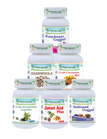

Product description:
1. Kanchnaar Guggul
Kanchnaar Guggul is an Ayurvedic polyherbal formulation containing Kanchnaar (Bauhinia variegata), Amalaki, Haritaki, Bibhitaki, Trikatu, Elaichi, Cinnamon, Tejpatra, Varuna bark, and Guggul. In the Ayurvedic management of PVNS, it may be considered as a supportive formulation when features of localized tissue accumulation, Kapha aggravation, and Srotas dysfunction are present. Kanchnaar and Guggul are traditionally valued for their Lekhana and Granthi-hara properties, while Triphala supports metabolic balance and Trikatu promotes Deepana-Pachana. The aromatic herbs further support digestion and circulation. Its use should be individualized according to Dosha, Agni, disease stage, and patient strength
Dosage: 2 tablets twice a day with warm water.
2. Crab Curcumin 95%
Crab Curcumin 95%, containing Haridra (Curcuma longa) and Maricha (Piper nigrum), may be considered as a supportive Ayurvedic formulation in the management of Pigmented Villonodular Synovitis. Haridra is recognized for its Shothahara (anti-inflammatory) and Rasayana properties and contains curcuminoids, while Maricha is valued for its Deepana-Pachana actions and may support the bioavailability of curcumin. In an Ayurvedic approach, these properties may be useful for supporting management of joint inflammation, discomfort, and functional stiffness associated with the condition.
Dosage: 1 capsule twice a day after meals with plain water.
3. Dashmoola Capsules
Dashmoola Capsules contain a combination of Ayurvedic herbs including Kantakari (Solanum surattense), Brihati (Solanum indicum), Bilwa (Aegle marmelos), Agnimantha (Clerodendrum phlomidis), Gokshura (Tribulus terrestris), Shalparni (Desmodium gangeticum), and Gambhari (Gmelina arborea). In the supportive management of Pigmented Villonodular Synovitis, this formulation may be considered for its Shothahara (anti-inflammatory), Vedanasthapana (pain-relieving), and Vata-balancing properties. The constituent herbs may help support comfort, mobility, and functional recovery in patients experiencing joint pain and stiffness.
Dosage: 1 capsule twice a day after meals with plain water.
4. Crab Artemisinin
Crab Artemisinin, containing Davana extract and Maricha extract, may be considered as a supportive formulation in the Ayurvedic management of Pigmented Villonodular Synovitis (PVNS). Davana is traditionally valued for its Vata-Kapha balancing and Shothahara (anti-inflammatory) properties, while Maricha (Piper nigrum) possesses Deepana-Pachana actions and supports the assimilation of herbal constituents. Together, these ingredients may provide supportive care for symptoms such as joint discomfort, stiffness, and localized inflammatory features associated with PVNS.
Dosage: 1 capsule twice a day after meals with plain water.
5. Joint Aid Plus
Joint Aid Plus, containing Nirgundi (Vitex negundo), Shallaki (Boswellia serrata), Guggul (Commiphora mukul), Sonth (Zingiber officinale), and Ashwagandha (Withania somnifera), may be used as a supportive formulation for joint-related symptoms associated with Pigmented Villonodular Synovitis (PVNS). Nirgundi and Shallaki are traditionally recognized for their Shothahara (anti-inflammatory) and pain-supportive properties, while Guggul is valued for its Vata-Kapha balancing action. Sonth supports Deepana-Pachana and may help address stiffness and impaired joint comfort, whereas Ashwagandha provides Balya and Rasayana support for musculoskeletal function. Collectively, the formulation may support reduction of joint discomfort, stiffness, and functional limitations.
Dosage: 1 capsule twice a day after meals with plain water.
6. Sinhnaad Guggul
Sinhnaad Guggul, also referred to as Simhanad Guggul, contains Haritaki, Bibhitaki, Amalaki, Shuddha Gandhak, Shuddha Guggul, and Erand oil. In the supportive Ayurvedic management of Pigmented Villonodular Synovitis (PVNS), this formulation may be considered when joint pain, stiffness, and inflammatory features are prominent. Triphala provides a combination of Deepana-Pachana and Rasayana support, while Shuddha Guggul is traditionally used for disorders involving Vata and Kapha and localized inflammatory processes. Erand oil contributes Vata-anulomana and Shothahara support, whereas Shuddha Gandhak is traditionally regarded for its supportive effect on tissue metabolism.
Dosage: 2 tablets twice a day with warm water.
Importance of Yoga and Local Basti in Pigmented Villonodular Synovitis
From an Ayurvedic perspective, supportive care in Pigmented Villonodular Synovitis (PVNS) should focus on maintaining joint mobility, reducing functional stiffness, and supporting comfort. Yoga and appropriately selected local Basti therapies may be incorporated as complementary measures, particularly during rehabilitation.
Role of Yoga
- Maintaining joint mobility: Gentle yoga movements can help preserve the range of motion of an affected joint and reduce stiffness associated with prolonged inactivity.
- Improving flexibility: Controlled stretching may maintain flexibility of the muscles, tendons, and periarticular tissues surrounding the affected joint.
- Supporting muscle strength: Appropriate strengthening practices can help maintain muscular support around the involved joint, which may improve functional stability.
- Reducing mechanical stress: Better flexibility, muscle conditioning, and body awareness can help patients perform daily movements with less unnecessary loading of the affected joint.
- Improving circulation and relaxation: Gentle movement and regulated breathing may support general circulation and relaxation, which can be useful for patients experiencing chronic musculoskeletal discomfort.
Role of Local Basti
Local Basti, such as Kati Basti, Janu Basti, or Greeva Basti, involves retaining appropriately selected warm medicated oil or other prescribed material over a localized body region for a defined period. The specific Basti is selected according to the anatomical site and clinical presentation.
- Janu Basti: May be considered as supportive therapy when the knee is affected, particularly for localized stiffness and discomfort.
- Kati Basti: May be considered when symptoms involve the lumbar or lumbosacral region, although it does not directly treat an intra-articular PVNS lesion.
- Greeva Basti: May have a supportive role when the cervical region is involved and stiffness or discomfort is present.
- Vata-related symptoms: Warm local therapies are traditionally used to provide Snehana and Swedana-like effects, which may be useful when stiffness and pain are prominent.
- Local comfort: The warmth and oil application may temporarily improve subjective comfort and ease of movement in some patients.
- Musculoskeletal support: Local Basti can be incorporated into an individualized Ayurvedic rehabilitation plan alongside appropriate exercise, physiotherapy, and lifestyle measures.
Conclusion
Effective management of Pigmented Villonodular Synovitis requires timely recognition, appropriate assessment, and an individualized therapeutic approach. Early intervention can help preserve joint function, minimize structural damage, and improve quality of life. Surgical management remains important for controlling localized or extensive disease, while targeted therapies may provide additional options in selected cases. Long-term follow-up is valuable because recurrence can occur, particularly with diffuse involvement. Supportive measures such as physiotherapy, appropriate physical activity, and symptom management can contribute to functional recovery.


Dr. Vikram Chauhan
Latest posts by Dr. Vikram Chauhan (see all)
- Pigmented Villonodular Synovitis (PVNS): Symptoms, Causes & Treatment - September 25, 2026
- Idiopathic Thrombocytopenic Purpura (ITP): Causes, Symptoms, Treatment & Ayurveda - September 24, 2026
- Ulcerative Colitis: Symptoms, Causes & Ayurvedic Treatment - September 23, 2026
- Fatty Liver: Causes, Symptoms, Diet & Ayurvedic Management - September 22, 2026
- Ulcerative Colitis: Symptoms, Causes With Ayurvedic Treatment - September 21, 2026