


Cavities Explained – Symptoms, Stages, Treatment & Prevention
Abstract
According to ancient texts, the main cause of dental cavities was a ‘tooth worm’. Caries is a Latin word that means ‘decay’. Caries is one of the oldest and most common problems in humans. It is a very prevalent disease globally. The bacteria causing caries (cariogenic bacteria) reside in the mouth and feed on acidic, sugary, and starchy food particles. It mostly affects people who frequently consume sugar. The site of development of a carious lesion varies. It can affect any of the tooth surfaces. Early lesions are usually not visible, but later stages present with certain signs and symptoms. Caries must be treated on time, and proper preventive measures must be taken to prevent further decay.
Statistics for Dental Caries
- Around 48% of people globally are prone to dental caries.
- 62% of the population above 18 years is affected, and about 52% is 3–18 years of age.
- 85% of senior people are affected by dental caries.
- 13.2% of children aged 5–19 years have untreated carious lesions.
- 20.2% of adults aged 65 years and above have untreated caries.
Introduction
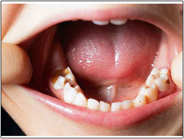
Caries is a chronic disease that results from cariogenic bacteria, most commonly Streptococcus mutans, that metabolise sugars and produce acids. The acid production causes demineralisation of the tooth’s structure. The carious lesion occurs in the dental hard tissues and causes fluctuations in the pH. It is a multifactorial disease. Various factors play a major role in its aetiology, such as bacteria, diet, behavioural, social, and psychological factors. Caries is also referred to as a dietary-bacterial disease because the bacteria feed upon the food debris stuck in the teeth. Fermentable carbohydrates such as glucose, sucrose, fructose, or maltose become the substrate for the cariogenic bacteria.
According to the modern concept of caries, there are four factors involved in caries formation.
These include
- Plaque
- Substrate
- Susceptible tooth
- Time
The presence of the above factors simultaneously causes demineralisation. If any of the factors are missing, the demineralisation process will not start.
The risk factors associated with caries formation include
- Diet
- Fluoride exposure
- Oral hygiene
- Xerostomia (dry mouth)
- Age and sex
The location of the carious lesion can vary. Caries on the crown part of the tooth are called coronal caries, whereas caries on the root are called radicular caries.
According to G.V. Black, caries is classified into six classes:
- Class 1 – It includes pit and fissure caries.
- Class 2 – It includes caries on the mesial or distal surfaces of posterior teeth (molars and premolars).
- Class 3 – It includes caries on the incisal edge of the anterior teeth (incisors and canines).
- Class 4 – It includes caries on the anterior teeth, excluding the incisal edge.
- Class 5 – It includes caries on the gingival one-third of the teeth on the lingual and facial surfaces.
- Class 6 – It includes carious lesions on the cusp tips of the posterior teeth.
Signs and Symptoms of Dental Caries
The signs and symptoms may vary in each individual and depend on the location on the tooth’s surface. Sometimes, early signs are not visible, and the caries progress towards advanced stages. At this stage, signs and symptoms occur that need to be treated. Some of the signs and symptoms include:
- Sensitivity of the tooth
- White or brown spots
- Pits or holes
- Staining or discolouration of the tooth
- Bad breath
- Mild to moderate pain upon mastication
- Gross decay in advanced stages
- Swelling or pus formation in advanced stages
Stages of Caries Progression
The signs and symptoms may vary in each individual and depend on the location of the lesion on the tooth surface. Sometimes, early signs are not visible, and the caries progresses towards advanced stages. At this stage, signs and symptoms occur that need to be treated. Some of the signs and symptoms include:
Stage 1: Initial stage (Demineralisation)
Enamel is the outermost and hardest layer of the tooth, which is made up of minerals. It is more prone to attack by acids. With prolonged exposure to acids, enamel loses its mineral content, and chalky white spots appear. This process is called demineralisation. It is counteracted by the body’s defence mechanism, and remineralisation occurs. Fluoride is a mineral that is required for the remineralisation process. If remineralisation fails to occur at this stage, the caries progresses to the second stage. These chalky white spots can be missed during examination as they may resemble the tooth’s natural colour.
Stage 2: Decay of Enamel
When acid exposure continues for a longer period, the remineralisation ability declines, and the enamel surface continues to demineralise further. The chalky white spots start to become brownish-black in colour. There are no symptoms of pain or sensitivity at this stage. The hydroxyapatite crystals of the enamel are broken down by organic acids.
Stage 3: Decay of Dentin
With continuous breakdown of the enamel, the lesion enters the inner soft layer called dentin. The dentin layer has dentinal tubules that contain dentinal fluid. This fluid transmits pain sensations to the nerves of the tooth. At this stage, sensitivity is felt upon mastication or exposure to hot or cold foods. Mild pain can also be felt that may linger for a few seconds. The decay progresses faster once it reaches the dentinal layer.
Stage 4: Pulpal tissue damage
The next or advanced stage of decay occurs when the pulp becomes involved in the process. The pulp is the innermost layer of the tooth that contains nerves and nerve fibres. At this stage, moderate to severe pain is felt. The pain may be continuous, throbbing, sharp, and may linger for about 30 seconds. Discomfort and sensitivity increase further. The pulp tissue becomes inflamed, leading to pulpitis, which can be reversible or irreversible depending on the symptoms. It requires urgent care and treatment.
Stage 5: Abscess formation
This is the last stage of decay. Swelling and infection occur with pus formation. With untreated caries, abscess formation starts. This causes severe pain. The pain may be radiating in nature. It can radiate to the jaw, gums, face, and even involve the lymph nodes. The infection reaches the apex of the tooth and then crosses the apex. Immediate treatment is required to drain the abscess and perform endodontic therapy.
Treatment and Prevention Strategies for Dental Caries
The treatment of caries depends on the severity and stage of progression. Early carious lesions are difficult to see with the naked eye but can be detected upon examination. The soft carious lesion gives a ‘catch’ with the explorer. It can be excavated easily and gives a good prognosis. Advanced lesions can lead to gross decay and involvement of deeper layers of the tooth structure.
The Treatment Methods Include
- Fluoride application: Fluoride helps in remineralising the enamel surface and can reverse the early stage of caries. Fluoride is present in tap water, mouthwashes, and toothpaste. The use of fluoridated toothpaste and mouthwashes can be helpful. Topical fluoride is applied by the dentist in the form of foam, gel, or varnish. These are brushed onto the tooth surface or placed in a tray that is fitted over the teeth.
- Restorations: In this method of treatment, different materials are used to restore the carious lesion. Restorations are helpful when the pulp has not been reached. The carious lesion is excavated with an air rotor, and a cavity is prepared with well-defined walls and margins. The filling material is then placed into the prepared cavity. The filling materials can be tooth-coloured, such as composite or GIC, or silver-coloured, such as amalgam. These materials are biocompatible and do not irritate the tissues. The restoration is then cured with a curing light, and the occlusal surface is shaped to resemble the natural tooth with grooves and fissures.
- Root canal treatment: When the tooth becomes sensitive to external stimuli such as hot and cold stimuli, it is indicative of irreversible pulpitis. Pain is sharp, throbbing, and lingers for a few seconds. The pulp tissue is either affected or becomes necrosed. In cases of abscess formation, root canal treatment is required with prior drainage. The patient is also given antibiotic prophylaxis before treatment. In this procedure, the damaged pulp tissue is removed, the canals are biochemically irrigated and obturated with root canal filling materials. Root canal therapy is followed by the placement of a dental crown to support the tooth structure and provide strength.
- Extraction: In some cases, the tooth becomes grossly decayed with little to no structure left behind. These cases are managed by tooth extraction, and the missing tooth is replaced with an artificial tooth to maintain occlusion and aesthetics. Dental implants can also be placed at the missing tooth site.
The Key Preventive Measures for Dental Caries Include
- Good oral hygiene: Brushing the teeth twice a day with fluoridated toothpaste is important to prevent future caries. Proper brushing technique must be followed. Daily flossing helps remove trapped food particles between the teeth. Rinsing with an alcohol-free mouthwash is also an effective strategy.
- Diet and lifestyle: Limit the consumption of sugary, sticky, or acidic foods and drinks. These become food for the cariogenic bacteria in the oral cavity. The bacteria feed upon fermentable carbohydrates and produce acids that attack the enamel, causing demineralisation. Quit smoking and avoid the use of tobacco products.
- Hydration: Drink plenty of water daily. This helps keep the mouth moist and flushes away bacteria. Insufficient water intake can cause xerostomia or dry mouth. With reduced salivary flow, bacteria thrive in the oral cavity and cause damage.
- Dental sealants: Sealants are applied to the biting or occlusal surfaces of the teeth. These are thin protective coatings that prevent food debris from getting stuck in the grooves of the teeth.
- Regular dental visits: Visiting the dentist every six months for routine checkups is important in preventing the disease. A thorough examination and early detection of carious lesions can prevent further decay and protect the tooth structure.
Conclusion
Dental caries is one of the most common problems faced by children, teenagers, and older adults. It presents with different symptoms depending on whether it is an early or advanced lesion. Various factors are involved in its aetiology, including bacteria, diet, susceptible tooth, age, and gender. The location of the lesion can be on the mesial, distal, occlusal, or lingual surfaces of the tooth. There are different stages of caries progression. Depending on the severity, appropriate treatment is provided. As prevention is better than cure, regular dental visits should be made to diagnose caries early through proper examination.


Dr. Vikram Chauhan
Latest posts by Dr. Vikram Chauhan (see all)
- Hemangiomas(benign vascular tumors) , Causes, Symptoms, Key Facts & Treatment - October 16, 2026
- Top 6 Herbs for stronger bones as per Ayurvedic Texts - October 12, 2026
- Top 4 Herbs for Stronger Liver as per Ayurvedic Texts - October 6, 2026
- Management of Olfactory neuroblastoma (ONB) in Ayurveda - September 28, 2026
- What is Aroma Therapy? – How is it an important part of Indian and different cultures since long? - September 18, 2026