


Erythema Dyschromicum Perstans: Causes, Symptoms & Treatment
Abstract
Pigmentary skin disorders are a common concern due to their impact on appearance and psychological well-being. Erythema Dyschromicum Perstans (EDP) is a rare, chronic skin disorder characterized by slowly progressive ash-gray or blue-gray patches, most commonly affecting the trunk, neck, face, and upper limbs. The condition is generally asymptomatic, although mild itching may occur in some individuals. Owing to its characteristic gray discoloration, EDP is widely known as Ashy Dermatosis and is also referred to as Dermatosis Cenicienta (“Cinderella Dermatosis” or “Ash-colored Dermatosis”) in Spanish-speaking regions. The exact cause remains unclear, and the disease can be difficult to diagnose because it resembles several other pigmentary disorders.
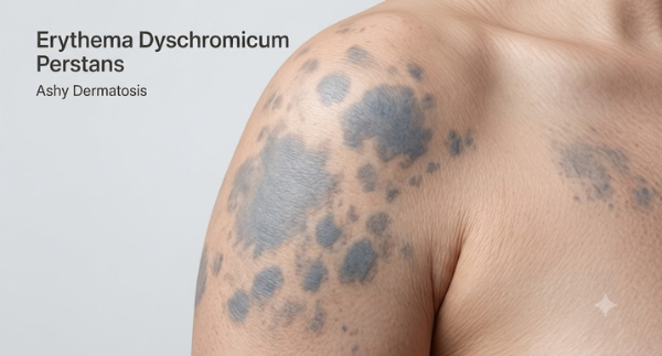
Statistics for Erythema Dyschromicum Perstans
- EDP is observed more frequently in females. Recent clinical studies have reported that nearly 94% of diagnosed cases occur in women.
- The condition is most commonly diagnosed in young adults, with an average age of around 26 years.
- EDP is reported more often among Hispanic, Latino, and Asian populations.
- Approximately one-third of individuals with EDP have one or more accompanying autoimmune diseases, suggesting a possible link between immune system dysfunction and the condition.
Facts about Erythema Dyschromicum Perstans
- Also known as Ashy Dermatosis: Erythema Dyschromicum Perstans (EDP) is commonly called Ashy Dermatosis because of its characteristic ash-gray to blue-gray skin patches.
- Rare pigmentary disorder: EDP is an uncommon skin condition, making it one of the less frequently encountered causes of acquired skin hyperpigmentation.
- Most patients are diagnosed in their twenties, although the condition can occur at any age, including childhood.
- EDP is often confused with lichen planus pigmentosus, post-inflammatory hyperpigmentation, fixed drug eruption, and other causes of acquired pigmentation, making accurate diagnosis important.
- EDP cannot spread from one person to another through physical contact.
- No risk of skin cancer: The condition affects skin pigmentation but is not considered a precancerous disorder and does not increase the risk of skin cancer.
Introduction
Erythema Dyschromicum Perstans (EDP) is a rare, acquired pigmentary skin disorder characterized by the gradual appearance of ash-gray, blue-gray, or slate-colored patches on the skin.
Common sites where lesions occur are:
- Trunk
- Neck
- Face
- Upper limbs
EDP is widely known as Ashy Dermatosis, a name that reflects the distinctive ash-colored appearance of the skin lesions. In Spanish-speaking countries, it is also referred to as Dermatosis Cenicienta. The word cenicienta means ash–colored and is also the Spanish name for the fairy-tale character Cinderella. For this reason, the condition is sometimes informally translated as “Cinderella Dermatosis.” However, the term actually refers to the ash-gray pigmentation of the skin rather than having any association with the fairy tale itself. The terms Erythema Dyschromicum Perstans and Ashy Dermatosis are often used interchangeably in medical literature. EDP is reported worldwide but is seen more frequently among individuals of Hispanic, Latino, Asian, and darker skin phototypes. It is more common in females and is typically diagnosed in young adults, although children and older adults may also be affected. The condition is benign and non-contagious, but its visible skin discoloration can significantly affect self-confidence, emotional well-being, and quality of life.
Etiological Factors of Erythema Dyschromicum Perstans
The exact cause of Erythema Dyschromicum Perstans (EDP), also known as Ashy Dermatosis, has not yet been established. The condition is considered idiopathic, meaning that no single definitive cause has been identified. Current evidence suggests that EDP is likely the result of a cell-mediated immune response, in which immune cells mistakenly target the pigment-producing cells (melanocytes) or the basal layer of the skin. This process leads to pigment leakage into the deeper layers of the skin, resulting in the characteristic ash-gray discoloration. Several factors have been proposed as possible triggers or contributors to the development of EDP. These include:
Genetic predisposition
Certain individuals may have an inherited susceptibility to developing EDP. There is an association with specific Human Leukocyte Antigen (HLA) types, particularly HLA-DR4, suggesting that genetic factors may influence the body’s immune response.
Immune system abnormalities
Activated T-lymphocytes may damage basal keratinocytes and melanocytes, leading to pigment incontinence and persistent grayish pigmentation. This immune-mediated process may also explain the association of EDP with autoimmune disorders in some patients
Exposure to chemicals
Contact with or ingestion of certain chemical substances has been suggested as a possible trigger. EDP is also linked with exposure to ammonium nitrate, commonly used in fertilizers, as well as barium sulfate.
Infections
These include intestinal parasitic infections, particularly those caused by nematodes, as well as certain viral infections. Chronic infections such as hepatitis C and human immunodeficiency virus (HIV) have also been reported in association with EDP.
Medications
Medications that have been associated with the condition include penicillin, benzodiazepines, omeprazole, and epidermal growth factor receptor (EGFR) inhibitors used in cancer therapy. The skin pigmentation may appear after prolonged use or as an uncommon adverse drug reaction.
Vaccination
Rare cases of EDP have been described following immunization, including influenza and COVID–19 vaccines. It is believed that vaccination may act as a trigger in susceptible individuals rather than directly causing the disease.
Contact allergy
Allergic reactions to certain cosmetics, fragrances, hair dyes, and other topical products are possible initiating factors. Persistent skin inflammation caused by allergic contact dermatitis may contribute to pigmentary changes in predisposed individuals.
Environmental and occupational factors
Repeated exposure to environmental allergens, industrial chemicals, or occupational irritants may contribute to disease development in genetically susceptible individuals.
Clinical Features of Erythema Dyschromicum Perstans
Most patients remain free from significant physical symptoms, making the condition mainly a cosmetic concern rather than a serious health problem. Recognizing its characteristic appearance and distribution is important for differentiating EDP from other pigmentary disorders and establishing an accurate diagnosis. The main features are:
Characteristic skin discoloration
The hallmark feature of EDP is the appearance of persistent ash-gray, blue-gray, slate-gray, or bluish-brown macules and patches. The pigmentation gradually becomes more noticeable over time and often remains for several months or years.
Early inflammatory border
In the initial stage, newly developing lesions may be surrounded by a slightly elevated erythematous (reddish) border. This active margin represents the inflammatory phase of the disease. As the lesions mature, the redness gradually disappears, leaving behind flat, grayish pigmentation.
Variable shape and size
The lesions may be round, oval, or irregular in shape. Their size varies considerably, ranging from approximately 0.5 cm to several centimeters in diameter. Over time, neighboring lesions may enlarge and merge, forming larger areas of pigmentation.
Symmetrical distribution
EDP most commonly affects the trunk, particularly the chest and back, followed by the neck, face, upper arms, and proximal lower limbs. The lesions are usually distributed symmetrically, involving similar areas on both sides of the body.
Predominance in darker skin phototypes
Although EDP can occur in individuals of any ethnicity or skin color, it is reported more frequently in people with medium to dark skin tones, where the grayish pigmentation is often more apparent.
Absence of significant symptoms
Most patients do not experience pain, burning, tenderness, or other physical discomfort. In many cases, the condition is discovered solely because of the visible skin discoloration.
Occasional mild itching
A small proportion of individuals may experience mild pruritus (itching), particularly during the early inflammatory stage when the reddish border is still present. Persistent or severe itching is uncommon.
No systemic manifestations
EDP is primarily confined to the skin and does not affect internal organs or overall physical health. It is not associated with fever, weakness, fatigue, weight loss, or other systemic symptoms.
Chronic but benign course
The condition usually progresses slowly and follows a chronic course. Although the pigmentation may persist for years, EDP is considered a benign, non-contagious disorder and does not increase the risk of skin cancer or other serious medical complications.
Diagnosis of Erythema Dyschromicum Perstans
The diagnosis of Erythema Dyschromicum Perstans (EDP) is mainly based on a detailed medical history, careful clinical examination, and characteristic skin findings. Since there is no single laboratory test that confirms the disease, dermatologists rely on the combination of clinical features and histopathological examination when necessary. The diagnosis include:
Clinical Evaluation
- Characteristic skin lesions – Presence of ash-gray, slate-gray, bluish-gray, or grayish-brown macules and patches.
- Lesions usually develop gradually and enlarge slowly over time.
- The pigmentation is persistent and often remains for months or years.
- Newly formed lesions may show a slightly raised reddish (erythematous) border.
- As the lesion matures, the red margin disappears, leaving a flat gray patch.
- Lesions commonly involve the trunk, especially the chest and back. Other frequently affected sites include the neck, upper arms, shoulders, and proximal lower limbs.
- Most patients have no pain, burning, or tenderness.
- Mild itching may occur in a few individuals, particularly during the early inflammatory stage.
- The condition does not produce fever, weight loss, fatigue, or other systemic symptoms.
Medical History
A detailed medical history helps identify possible triggering factors and exclude other causes of skin pigmentation. The physician may ask about:
- Duration and progression of the lesions
- Previous episodes of skin inflammation or rashes
- Current and past medications
- History of allergies or chemical contact
- Recent infections
- Family history of pigmentary disorders
- Presence of autoimmune diseases or other chronic illnesses
Skin biopsy
A skin biopsy is often recommended when the diagnosis is uncertain or when other pigmentary disorders need to be excluded. During this procedure, a small sample of affected skin is removed under local anesthesia and examined microscopically.
Histopathological findings
Microscopic examination of the skin may reveal several characteristic changes:- Degeneration or vacuolar alteration of the basal cell layer of the epidermis is commonly observed.
- A mild lymphocytic infiltrate is usually present around superficial dermal blood vessels.
- Melanin pigment leaks from the damaged basal layer into the upper dermis.
- Numerous melanophages (macrophages containing melanin pigment) are commonly seen in the superficial dermis.
- Occasionally, small deposits of hemosiderin may also be observed.
Management of Erythema Dyschromicum Perstans
Treatment is generally selected according to the severity of the condition, whether active inflammation is present, and how much the pigmentation affects the patient. Some therapies aim to reduce inflammation and prevent the development of new lesions, while others are used to improve persistent pigmentation. The treatment options include:
Topical corticosteroids
Mild to moderate topical corticosteroids may be prescribed for a limited period when newly developing lesions show redness or inflammation. They are mainly intended to control the inflammatory component of EDP rather than directly remove established pigmentation. Prolonged or inappropriate use should be avoided because topical steroids can cause skin thinning and other adverse effects.
Topical calcineurin inhibitors
Medicines such as tacrolimus ointment may be considered when inflammation needs to be controlled, particularly in areas where long-term steroid use is undesirable. These medications can help reduce inflammatory activity without causing the skin thinning associated with prolonged corticosteroid use.
Narrow band UVB phototherapy
It may help reduce active inflammatory changes and improve pigmentation in selected cases. Treatment usually requires repeated sessions under dermatological supervision, and the response is variable.
Clofazimine
Clofazimine is an oral medication that has shown beneficial results in some published reports and is considered one of the systemic options for difficult cases. However, it can produce reddish-brown or darker skin discoloration and other adverse effects.
Dapsone
Dapsone has also been used as a systemic treatment, particularly when active inflammatory lesions continue to develop. Some patients may experience improvement while taking the medicine, although pigmentation can persist or return after treatment is discontinued. Dapsone requires appropriate medical monitoring because it can cause significant adverse effects in susceptible individuals.
Laser treatment
Pigment-targeting lasers, including Q-switched laser systems, have occasionally been used to address persistent pigmentation. Results are not consistent, and there is a possibility of unwanted changes in skin color, particularly in individuals with darker skin tones.
Chemical peels
Certain chemical peels have been attempted to improve stubborn pigmentation. However, excessive irritation or inflammation may sometimes worsen pigmentation or produce uneven skin color. For this reason, peels should only be considered after careful assessment by a dermatology professional.
Ayurveda Insights
The clinical presentation of Erthema Dischromicum Perstans may be considered in relation to disorders involving Twak (skin) and abnormal pigmentation, with possible involvement of Pitta and Vata doshas. Ayurvedic management may focus on balancing doshas, supporting healthy skin function, improving digestion and metabolism, and using individualized herbal approaches under qualified supervision.
Pathogenesis of Erthema Dyschromicum Perstans
The grayish skin discoloration in EDP may be understood conceptually through disorders of Twak (skin), Rakta (blood), and Pitta, with participation of Vata. Aggravated Pitta may disturb the normal coloration and metabolism of the skin, while Vata can contribute to dryness and irregular progression. Impaired Rakta Dhatu and Rasavaha/Raktavaha Srotas may further disturb skin nourishment and pigmentation. Accumulation of Ama due to impaired Agni may act as an additional contributing factor. Thus, the condition may be interpreted as a multifactorial disturbance involving Dosha imbalance, Dhatu dysfunction, and impaired skin metabolism.
Herbal Remedies for Erythema Dyschromicum Perstans by Planet Ayurveda
Planet Ayurveda is a well-established Ayurvedic healthcare organization that works to promote the ancient knowledge of Ayurveda through natural remedies and holistic health solutions. The company develops a wide range of herbal formulations using pure, high-quality ingredients and standardized herbal extracts, with a focus on maintaining safety, authenticity, and effectiveness. Following the principles of classical Ayurveda, Planet Ayurveda aims to support overall health by addressing the underlying causes of health concerns and encouraging balance in the body, mind, and emotions. With an emphasis on quality, natural ingredients, and traditional approaches supported by modern research, the organization works toward making Ayurvedic wellness solutions available globally. We will explore various herbal approaches used for the management of Erythema Dyschromicum Perstans according to Ayurvedic principles. The herbal formulations are:
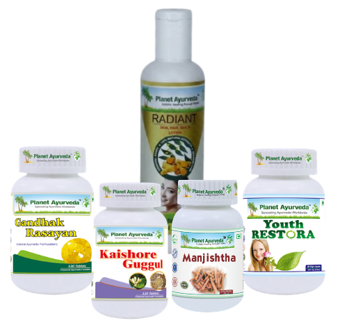
- Gandhak Rasayan
- Kaishore guggul
- Radiant skin, hair and nails lotion
- Youth Restora capsules
- Manjishtha capsules
Herbal Remedies for Erythema Dyschromicum Perstans

Product Discription
1. Gandhak Rasayan
Gandhak Rasayan, containing purified sulfur (Shuddha Gandhak), is traditionally used in Ayurveda for various skin disorders. In the Ayurvedic approach to Erythema Dyschromicum Perstans (EDP), it may be considered for supporting Twak and Rakta Dhatu and maintaining healthy skin function. Sulfur has documented antimicrobial and anti-inflammatory properties, although these effects mainly relate to topical sulfur and do not establish Gandhak Rasayan as a treatment for EDP.
Dosage: 2 tablets twice a day with lukewarm water.
2. Kaishore Guggul
It is a classical Ayurvedic formulation containing Guggulu (Commiphora mukul), Triphala (Three myrobalans), Guduchi (Tinospora cordifolia), and other ingredients traditionally used in inflammatory and skin-related disorders. In the Ayurvedic management of EDP, it may be considered to support Pitta–Rakta balance, Agni, and healthy skin metabolism. Its proposed role is mainly supportive and aimed at reducing inflammatory tendencies and promoting proper tissue nourishment.
Dosage: 2 tablets twice a day with lukewarm water.
3. Radiant skin, hair and nails lotion
It is an herbal topical formulation containing ingredients such as curcumin, Neem (Azadirachta indica), jasmine, Guggul, sesame, and coconut. In the Ayurvedic management of EDP, it may be used as supportive external skin care to nourish and moisturize the affected skin. Its ingredients are traditionally associated with soothing and maintaining healthy skin.
Usage: Place 5-6 drops on your palm. apply on the affected area of your skin, scalp & nails twice daily, morning and night.
4. Youth Restora Capsules
Youth Restora is an Ayurvedic nutritional formulation containing Bhumi Amla (Phyllanthus niruri), Grape Seed, Go-Piyush (Bovine colostrum), and Amalaki (Emblica officinalis). In the supportive Ayurvedic management of EDP, it may help maintain healthy skin by providing antioxidant and nutritional support. Its ingredients may help address oxidative stress and support normal tissue nourishment and immune function.
Dosage: 2 capsules twice a day with plain water after meals.
5. Manjishtha capsules
Manjishtha Capsules, containing Rubia cordifolia, may be considered as supportive therapy in the Ayurvedic management of Erythema Dyschromicum Perstans (EDP). Manjishtha is traditionally described for Rakta Shodhana and skin disorders and may help support healthy skin and inflammatory balance. Modern research also reports antioxidant and anti-inflammatory properties of Rubia cordifolia.
Dosage: 2 capsules twice daily with plain water after meals.
Conclusion
Although its exact cause remains uncertain, immune-related mechanisms and several possible environmental or genetic factors may contribute to its development. Diagnosis depends mainly on clinical examination, supported by skin biopsy when necessary. There is no universally effective treatment, and outcomes vary among individuals. Early recognition, appropriate management, regular skin care, and medical supervision can help control active changes and address the cosmetic concerns associated with the condition.


Dr. Vikram Chauhan
Latest posts by Dr. Vikram Chauhan (see all)
- Depleted Mother Syndrome: Symptoms & Ayurvedic Care - October 24, 2026
- Ketogenic Diet Guide: Benefits, Foods and Risks - October 24, 2026
- Immune Thrombocytopenia (ITP): Symptoms, Causes & Treatment - October 23, 2026
- Atopic Dermatitis Treatment: Symptoms, Causes & Ayurveda - October 23, 2026
- Encapsulating Peritoneal Sclerosis: Causes & Treatment - October 22, 2026