


Herbal Remedies For Dog Ear Hematoma – Symptoms, Causes & Treatment in Ayurveda
Abstract
Dog’s ear hematoma, also called an auricular hematoma, is a condition in which blood accumulates between the skin and cartilage of the ear flap. It commonly develops after vigorous head shaking or repeated scratching, often associated with ear infections, allergies, parasites, foreign bodies, or trauma. The affected ear may appear swollen, warm, painful, and soft or fluctuant to touch. If left unmanaged, healing can result in thickened, wrinkled, or permanently deformed ear cartilage. Proper evaluation is important to identify and address the underlying cause while managing the hematoma itself. From an Ayurveda perspective, the condition can be viewed through the principles of Vata and Pitta involvement, particularly where pain, inflammation, swelling, and tissue disturbance are present. Ayurvedic supportive approaches may focus on reducing inflammation, promoting tissue healing, and addressing the underlying imbalance.
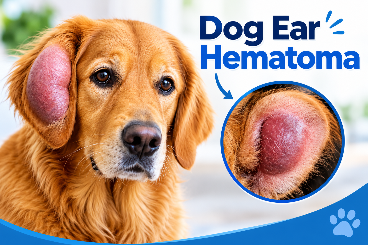
Statistics for Dog’s Ear Hematoma
- It affects approximately 0.25%–0.35% of dogs in primary veterinary care settings.
- It is more frequently observed in middle-aged and senior dogs, with some studies reporting higher occurrences between 8–12 years.
- The condition generally affects one ear at a time, although both ears can be involved in some dogs.
- Certain regional studies have reported a greater frequency of left-sided hematomas, reaching approximately 62% in specific groups.
- Around 45%–76% of affected dogs have underlying otitis externa, which can trigger persistent head shaking and scratching.
Facts about Dog’s Ear Hematoma
- A tiny blood vessel can create a surprisingly large swelling because the ear flap has limited space between its tissue layers.
- The ear flap can behave like a small “blood pocket,” allowing collected blood to spread across a wide area.
- An aural hematoma is not simply a bruise; it involves blood collecting between the layers of the ear flap.
- The final appearance of the ear can change permanently if the collected blood organizes and the tissue heals abnormally.
- A hematoma can feel different at different stages, becoming firmer as the accumulated blood undergoes organization.
- Dogs with floppy ears often attract special attention in veterinary practice because the ear flap has a broad surface area where hematomas can develop.
Introduction
A dog’s ears are delicate structures that contribute not only to hearing but also to communication, balance, and body language. Because the ear flap contains thin skin, cartilage, and numerous small blood vessels, it can be vulnerable to injuries and tissue damage. One important condition affecting this region is dog’s ear hematoma, medically known as an aural hematoma. It occurs when blood collects within the layers of the ear flap, creating a localized swelling that can vary considerably in size and appearance. An aural hematoma can develop suddenly, and pet owners may notice that the ear flap looks unusually enlarged or feels soft, fluid-filled, or thickened. Although it can appear alarming, the visible swelling represents only part of the condition. Every case is different. The size of the hematoma, its duration, the condition of the ear tissue, and the dog’s overall health all influence management and recovery.
Anatomy of the Dog’s Ear Flap
The ear flap, medically called the pinna or auricle, is the visible portion of a dog’s ear. It is a flexible, highly vascular structure designed to collect sound waves and direct them toward the ear canal. Its anatomy also explains why blood can accumulate within the flap and form an aural hematoma.
- Skin: The outer surface is covered by thin skin containing hair follicles, sebaceous glands, and a network of small blood vessels.
- Auricular cartilage: A flexible sheet of elastic cartilage forms the framework of the pinna and gives the ear its characteristic shape while allowing movement.
- Blood vessels: Numerous small arteries, veins, and capillaries run through the ear flap. Damage to these vessels can allow blood to collect within the tissue layers.
- Perichondrium: This thin connective-tissue layer surrounds the cartilage and provides structural support as well as an important route for nutrients reaching the cartilage.
- Subcutaneous tissue: A relatively thin layer of connective and fatty tissue lies between the skin and cartilage. Its thickness varies among breeds and individuals.
- Ear canal connection: The base of the pinna continues into the external acoustic meatus, forming the pathway through which collected sound reaches deeper parts of the ear.
- Nerve supply: Sensory nerves supply the pinna, making the ear responsive to touch, pressure, temperature, and injury.
How Does an Aural Hematoma Develop in Dogs?
An aural hematoma develops when small blood vessels within the dog’s ear flap become damaged, allowing blood to collect between the skin and the underlying cartilage. The pinna contains a thin layer of tissue closely attached to its cartilage, so bleeding in this confined space can gradually separate these layers and create a pocket of blood. Repeated movement of the ear can enlarge this space as fresh bleeding occurs. Initially, the collected blood remains relatively fluid, but with time it begins to clot and undergo tissue organization. The body then starts repairing the affected area, with inflammatory cells and connective tissue becoming involved in the healing process. As the hematoma matures, fibrosis and scar-tissue formation can make the ear flap thicker and less flexible. In some cases, the newly formed tissue contracts and pulls the cartilage into an altered shape.
Signs and Symptoms of Dog’s Ear Hematoma
The features can vary depending on the size and stage of the hematoma, but some signs are easy for pet owners to recognize during routine handling or grooming. These include:
- Visible ear swelling: The ear flap may become enlarged, with swelling affecting a small area or most of the pinna.
- Soft, fluid-filled swelling: A fresh hematoma often feels soft or fluctuant due to accumulated blood beneath the skin.
- Pain or tenderness: The dog may become uncomfortable when the affected ear is touched.
- Thickened ear flap: Older hematomas can become firm and cause noticeable thickening of the pinna.
- Drooping ear: The additional weight of the swelling may cause the ear to hang lower than usual.
- Ear scratching: The dog may scratch around or at the affected ear.
- Altered ear shape: Larger or longstanding hematomas can distort the normal contour of the pinna.
- Behavioral changes: Some dogs may become restless, less active, or reluctant to have their ears handled.
Diagnosis of Dog’s Ear Hematoma
Diagnosis is usually straightforward when the characteristic swelling of the ear flap is present, but veterinary examination is important to confirm the condition and identify associated ear problems.
- Ear examination: The external ear canal is inspected for inflammation, discharge, foreign material, or other abnormalities.
- Palpation: Gentle examination helps determine whether the swelling is soft, fluctuant, firm, or fluid-filled.
- Otoscopy: An otoscope may be used to obtain a clearer view of the ear canal and eardrum.
- Fine-needle aspiration: A small amount of fluid can be withdrawn to confirm that the swelling contains blood.
- Cytology: The collected material may be examined microscopically when infection or other abnormalities are suspected.
- Additional testing: Blood tests or imaging are generally considered when the clinical examination indicates a need for further investigation.
Conventional Veterinary Management for Dog’s Ear Hematoma
The management is selected according to the size, duration, severity, and recurrence of the hematoma, along with the condition of the ear canal. The management approaches include:
- Aspiration: A needle or catheter can be used to remove accumulated blood, particularly in selected small or early hematomas.
- Drain placement: A temporary drain can help prevent blood from accumulating again and allows continued drainage during healing.
- Surgical correction: Larger, chronic, or recurrent hematomas often require surgery to empty the blood pocket and re-establish contact between the tissue layers.
- Pain management: Appropriate veterinary analgesics are used to control discomfort during recovery.
- Managing ear disease: Concurrent otitis, allergy, parasitic infestation, or other identified ear disorders should be managed to reduce the risk of recurrence.
- Protective measures: An Elizabethan collar may be recommended to limit scratching and interference with the treated ear.
- Follow-up examination: Regular veterinary checks help assess healing, detect fluid reaccumulation, and determine when drains or dressings can be removed.
Ayurveda Insights
From an Ayurvedic perspective, a dog’s ear hematoma can be understood by considering the principles of Vata, Pitta, Rakta, and Mamsa Dhatu involved in localized tissue disturbance. The ear flap contains delicate tissues and blood vessels, and accumulation of blood can be viewed in relation to Rakta Dushti and disturbance of normal tissue circulation. Vata is associated with movement, pain, and tissue disruption, while Pitta relates to heat, inflammation, and changes involving Rakta. Ayurveda therefore emphasizes an individualized approach based on the animal’s overall condition and the stage of the lesion. Supportive care may focus on maintaining healthy tissue repair, appropriate nutrition, gentle local care, and reducing factors that interfere with healing.
Pathogenesis for Dog’s Ear Hematoma
In an Ayurvedic interpretation, the pathogenesis of a dog’s ear hematoma can be described as a local process involving Dosha aggravation, Rakta disturbance, and injury to the tissues of the Pinna. The process can be understood step by step rather than simply assigning the condition to one Dosha.
- Initial tissue disturbance: Repeated mechanical stress affecting the delicate structures of the ear flap can be viewed under Vata Prakopa, particularly because Vata governs movement, mobility, and physical disturbance within the body.
- Sira involvement: The small blood vessels (Sira) present within the ear flap may become injured. Ayurveda gives considerable importance to Sira as channels associated with the movement of Rakta Dhatu.
- Rakta accumulation: Following vascular disruption, blood becomes collected within the local tissue space. This can be conceptually correlated with abnormal localization of Rakta Dhatu, rather than its normal circulation through the body.
- Development of Shotha: Accumulation within the tissue produces localized Shotha, characterized by expansion and alteration of the affected area.
- Pitta-Rakta interaction: Because Pitta has an intimate relationship with Rakta, Pitta involvement can be considered when the local process includes inflammatory changes, increased warmth, or tissue irritation.
- Mamsa Dhatu involvement: The ear flap contains soft tissues surrounding the cartilage. Disturbance within these tissues can be viewed in relation to Mamsa Dhatu, particularly when prolonged swelling alters the normal texture and structure of the pinna.
- Srotodushti: Local disruption can interfere with the normal movement of nutrients, fluids, and metabolic products through the involved tissue channels. This can be conceptually described as Srotodushti, especially when obstruction and abnormal accumulation occur at the local level.
- Chronic tissue organization: When the accumulated material persists, the body gradually organizes and repairs the affected region. Fibrous tissue can develop, making the ear flap progressively thicker and less flexible.
Herbal Remedies for Dog’s Ear Hematoma by Planet Ayurveda
Planet Ayurveda is a well-established Ayurvedic healthcare organization that works to promote the ancient knowledge of Ayurveda through natural remedies and holistic health solutions. The company develops a wide range of herbal formulations using pure, high-quality ingredients and standardized herbal extracts, with a focus on maintaining safety,authenticity, and effectiveness. Following the principles of classical Ayurveda, Planet Ayurveda aims to support overall health by addressing the underlying causes of health concerns and encouraging balance in the body, mind, and emotions. With an emphasis on quality, natural ingredients, and traditional approaches supported by modern research, the organization works toward making Ayurvedic wellness solutions available globally. We will explore various herbal approaches used for the management of Dog’s Ear Hematoma according to Ayurvedic principles. The herbal formulations are:
- Inflam vet tablet
- Boswellia Curcumin
- Niere Vet Powder
- Punarnava Capsules
- Mahamanjishthadi Kwath
- Nirgujati Oil
Herbal Remedies for Dog Ear Hematoma
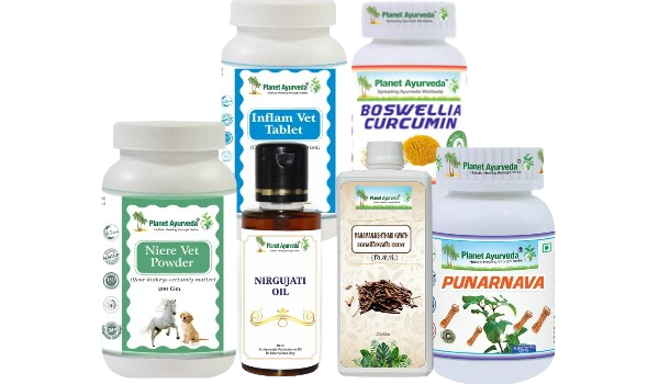

Product description:
1. Inflam Vet Tablets
These contain Shallaki (Boswellia serrata) and Guggul (Commiphora wightii), ingredients traditionally used for their anti-inflammatory properties. Shallaki may help reduce inflammatory responses and discomfort, while Guggul is used to support the management of inflammation. In dogs with an ear hematoma, these ingredients may serve as supportive care to help manage associated swelling and discomfort.
Dosage: 2 tablets twice a day after feed.
2. Boswellia Curcumin
It contains Shallaki (Boswellia serrata) and Haridra (Curcuma longa), two herbs known for their anti-inflammatory properties. Shallaki may help reduce inflammatory activity and support the management of discomfort, while Haridra contains curcumin, which helps regulate inflammatory responses. These ingredients may provide supportive care for dogs with ear hematoma by helping manage inflammation and associated tenderness.
Dosage: 1 capsule twice a day after feed.
3. Niere Vet Powder
It contains Varun (Crataeva nurvala), Gokshur (Tribulus terrestris), Kaasni (Cichorium intybus), Punarnava (Boerhavia diffusa), Bhumi Amla (Phyllanthus amarus), Shirish (Albizia lebbeck), Shigru (Moringa oleifera), and Apamarg (Achyranthes aspera). These herbs are traditionally used to support urinary and other physiological functions.
Dosage: 1 teaspoon twice a day after feed.
4. Punarnava Capsules
Punarnava (Boerhavia diffusa) is traditionally used for its diuretic, anti-inflammatory, and antioxidant properties. It supports urinary function and helps manage fluid retention. Its potential anti-inflammatory effects may offer supportive benefits in certain inflammatory conditions.
Dosage: 1 capsule twice a day after feed.
5. Mahamanjishthadi Kwath
It contains Manjistha (Rubia cordifolia), Kuda (Holarrhena pubescens), Nagarmotha (Cyperus rotundus), Sonth (Zingiber officinale), Neem (Azadirachta indica), Daruhaldi (Berberis aristata), Kutki (Picrorhiza kurroa), Shatavari (Asparagus racemosus), Varun (Crataeva nurvala), and various other herbs. These ingredients are traditionally used to support skin health and manage inflammatory conditions. Manjistha and Neem are valued for their potential anti-inflammatory properties, while Sonth contains compounds with anti- inflammatory activity. These properties may offer general supportive benefits.
Dosage: 10 ml to be taken twice a day after feed.
6. Nirgujati Oil
It contains Chamelipatra (Jasminum grandiflorum), Nimba Patra (Azadirachta indica), Haridra (Curcuma longa), Lodhra (Symplocos racemosa), Karanj Beej (Pongamia pinnata), Til Tail (Sesamum indicum), and Nirgundi (Vitex negundo) juice or decoction, along with other herbs. Several of these ingredients are traditionally valued for their skin-supporting and anti-inflammatory properties. Nirgundi and Haridra may help support the management of inflammation, while Neem is traditionally used in skin care.
Dosage: Apply it locally on the affected area.
Do’s and Don’ts for Pet Owners: Ayurvedic Perspective
- Do provide a simple, digestible diet during recovery to support normal digestion and tissue nourishment.
- Do maintain proper hydration to support overall recovery and normal physiological functions.
- Do keep the affected ear clean and protected according to veterinary advice.
- Do consider gentle supportive care only when specifically advised by a veterinary professional trained in Ayurveda.
- Don’t puncture, squeeze, or drain the hematoma at home.
- Don’t apply hot oils, herbal pastes, or irritant preparations directly to the swollen ear without professional advice.
- Don’t perform vigorous massage or rubbing over the hematoma.
- Don’t give strong Panchakarma procedures such as Vamana, Virechana, or Basti for this condition without specialized veterinary assessment.
- Don’t delay conventional treatment when drainage, surgery, pain control, or treatment of an underlying ear disorder is required.
Conclusion
Early veterinary evaluation is important to determine the extent of the hematoma and identify any associated ear problems. Appropriate management helps reduce discomfort, support healing, and preserve the natural shape of the pinna. From an Ayurvedic perspective, the condition can be viewed through concepts involving Vata, Pitta, Rakta Dhatu, Shotha, and local tissue disturbance. Ayurvedic measures can provide supportive care when selected appropriately.


Dr. Vikram Chauhan
Latest posts by Dr. Vikram Chauhan (see all)
- Herbal Remedies For Atopic Dermatitis – Symptoms, Causes & Treatment in Ayurveda - October 27, 2026
- Herbal Remedies For Dog Ear Hematoma – Symptoms, Causes & Treatment in Ayurveda - October 26, 2026
- Herbal Remedies for PCOD/PCOS: Symptoms, Causes & Ayurvedic Treatment - October 26, 2026
- Depleted Mother Syndrome: Symptoms & Ayurvedic Care - October 24, 2026
- Ketogenic Diet Guide: Benefits, Foods and Risks - October 24, 2026